Cone beam...What is it?
Cone beam computed tomography (CBCT) is a tomography technique that produces a digital x-ray.
On an X-ray, the bones appear white and the soft tissues in grey tones.
This technique, which emerged in the late 1990s, lies between the dental panoramic and the CT scanner and is creating a small revolution in the field of medical imaging by offering new opportunities for dental examination.
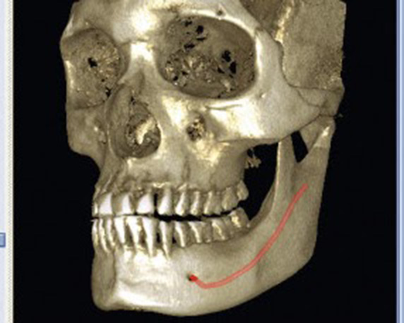
This device has the advantage of being more precise than the dental panoramic by offering a similar or even higher resolution than the scanner, with the added possibility of a 3D digital reconstruction. It allows an efficient examination of mineralized tissues (teeth, cartilage, bones), highlighting bone lesions, fractures, infections, cysts and foreign bodies with a very good accuracy.
Another advantage is that it can scan the entire volume to be radiographed in a single pass and is therefore much less irradiating than a scanner. It also offers the possibility of locating the examination field on the area to be studied (a few teeth, a jaw), which avoids unnecessary irradiation of other parts of the skull.
The cone beam is therefore the examination of choice for 3D imaging in dentistry.